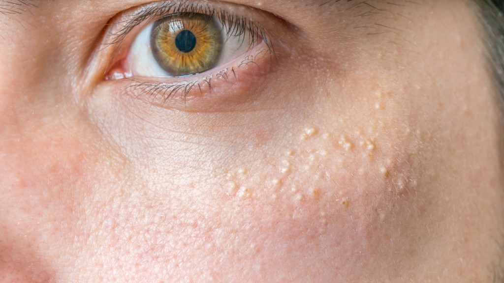
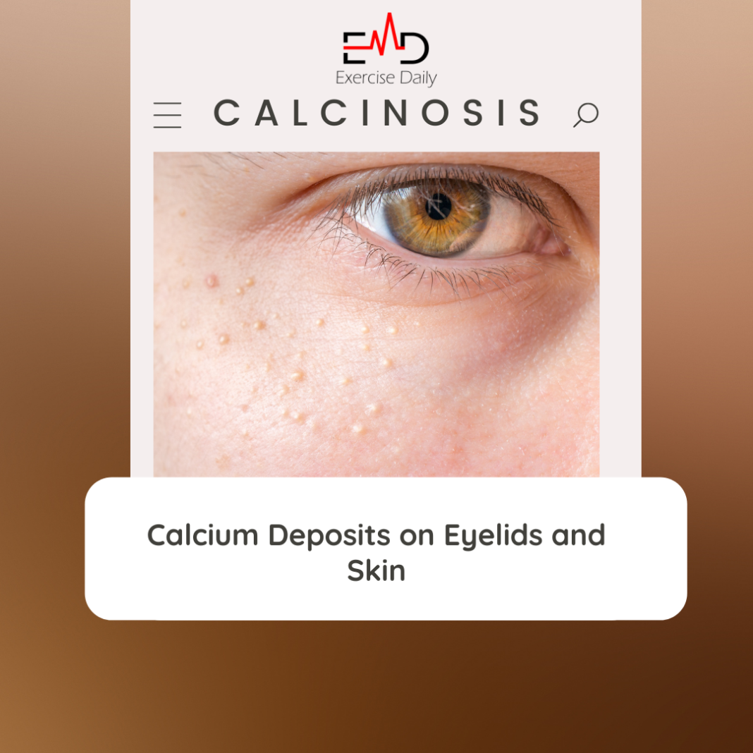
Exercise Daily – Calcinosis cutis is a term used to describe the formation of calcium deposits on the face. It might lead to calcium deposits on eyelids as well. Autoimmune diseases, acne, renal disease, and some high-dose calcium medicines are examples of such conditions.
Build up of calcium beneath the skin manifests itself as solid, white, or yellowish pimples on the skin surface. Hydroxyapatite is a mineral that your body utilizes to create and strengthen bones and teeth. Moreover, it is a kind of calcium phosphate that we often find in nature.
Calcification (also known as calcinosis) is a condition that arises when abnormally high levels of calcium phosphate accumulate in the soft tissues of the body. Calcinosis in the skin manifests as white or yellowish lumps on the skin’s surface.
Signs and Symptoms
Calcium deposits in the skin seem to appear out of nowhere on a regular basis. Those pimples might be a symptom or an indication of an underlying medical disease.

In the case of calcinosis, the most common symptom is the emergence of hard, pimple-like lumps or nodules. They are white or yellow in color. In addition, they possess the following characteristics:
- Bumps in variable sizes and amounts
- Often seen in groups
- If you punch a hole in this sort of nodule, a white, chalky, paste-like substance will flow out
- They might produce soreness and even pain in the region
- Bumps that appear around joints may cause joint stiffness
What is the Source of Calcium Deposits in the Skin?
Calcium deposits on eyelids and other body parts may be classified into four categories. Each of these is determined by the underlying cause of the condition:
- Dystrophic calcinosis cutis
- Iatrogenic calcinosis cutis
- Metastatic calcinosis cutis
- Idiopathic calcinosis cutis
Dystrophic calcinosis cutis
Dystrophic calcinosis may develop in tissue that has been injured or irritated. When injured tissues release proteins that bind calcium and phosphate, clumps of calcium and phosphate are formed that become larger and larger over time.
Moreover, it may also occur in a tissue that has turned cancerous or dead. The following are some of the conditions that might lead to dystrophic calcinosis cutis:
- Skin abrasion
- Infections of the skin
- Disorders of the connective tissue
- Panniculitis
- Acne
- Tumors
Iatrogenic calcinosis
Calcinosis caused by certain drugs and medical procedures, such as the frequent taking of blood from an infant’s heel, is sometimes referred to as iatrogenic calcinosis.
Metastatic calcinosis
Metastatic calcinosis may develop as a consequence of any medical disease that causes an excess of phosphorus or calcium.
When calcium or phosphate levels are elevated yet there is no tissue damage, this condition is known as hypercalcemia. The presence of high phosphate levels causes calcium to naturally bind to phosphate. These include the following:
- Calciphylaxis
- Kidney failure
- Sarcoidosis
- Paraneoplastic hypercalcemia
- Excess vitamin D
- Hyperparathyroidism
- Milk-alkali syndrome
Idiopathic calcinosis
Idiopathic calcinosis cutis is a kind of calcinosis that cannot be traced back to a particular etiology. However, it may cause calcium deposits on eyelids. The following are among the most common explanations that have been ruled out:
- The amounts of phosphorus and calcium in your body are within typical limits
- A prior injury to the tissue does not seem to have taken place
- You are not taking any drugs that might cause calcinosis
- You haven’t lately had any medical treatments that might have triggered calcinosis
Diagnosis
In order to determine if you have calcinosis cutis, your doctor will do a skin examination and evaluation of your medical history. If your calcium or phosphate levels are high, blood tests will be necessary to determine the cause.

Your doctor may recommend other blood tests which might help to determine if an underlying condition is present. The following tests may be necessary:
- Renal function tests (RFTs), which help to determine whether or not a patient has kidney disease.
- Check the level of parathyroid hormone to see whether you have hyperparathyroidism.
- Check for inflammation by measuring C-reactive protein (CRP) and erythrocyte sedimentation rate (ESR), which may occur with autoimmune illnesses, among other things.
- Calculated tomography (CT) scans and bone scanning may both be used to detect the extent of the calcium deposits in a patient’s bones.
In certain cases, people might misunderstand calcinosis cutis with other conditions such as milia (whiteheads) or gouty tophi (skin growths). Hence, a biopsy may confirm the diagnosis and rule out alternative possibilities.
What to Do if You Have Calcium Deposits on Eyelids and Skin?
Your doctor will discuss many therapies that are available to you and will suggest the one that they believe is the most appropriate for your case. Some of the alternatives are as follows:
- Intralesional corticosteroids (triamcinolone acetonide and triamcinolone diacetate)
- Calcium channel blockers, such as amlodipine (Norvasc), diltiazem (Cardizem, Tiazac), and verapamil (Calan, Verelan)
- Colchicine (Colcrys)
- Warfarin (Coumadin, Marevan)
- Laser therapy
- Iontophoresis
- Surgery to remove the calcium deposits
Iontophoresis involves the use of low levels of electric current to dissolve the calcium deposits by delivering medication. These medications include therapy with cortisone directly to the affected areas.
Non Conventional Treatments
When it comes to treating calcium deposits on the skin, there are a few natural methods you may try.
Massage
Many individuals believe that rubbing the afflicted region with aloe vera gel or olive oil helps in the eradication of calcium deposits.
Diet
Many natural healing proponents believe that reducing your calcium intake and eliminating foods such as dairy products may be beneficial.
Apple Cider Vinegar
Regularly consuming 1 tablespoon of apple cider vinegar combined with 8 ounces of water every day can aid in the breakdown of calcium deposits.
Chanca piedra
Another theory is that the herb chanca piedra might help to break down the accumulation of calcium in the bloodstream and bones. Hence, it might be beneficial for breaking calcium deposits on eyelids.
Frequently Asked Questions
How do you get rid of calcium deposits on your eyelids?
You can get rid of calcium deposits by mechanical debridement with a blade, chemical chelation with ethylenediaminetetraacetic acid (EDTA), and phototherapeutic keratectomy.
Manual debridement of CBK using a blade is successful. However, it might result in an uneven corneal surface as a result of the scraping motion.
What causes calcium deposits on eye lids?
Acne, skin infections, varicose veins, and burns are all possible causes. Moreover, certain autoimmune disorders such as lupus, rheumatoid arthritis, and scleroderma can also cause calcium deposits.
How do you get rid of calcium deposits?
- It may be necessary for a professional to numb the region and then use ultrasound imaging to guide needles to the deposit. It is necessary to loosen the deposit before he or she might draw out the majority of the deposit with the needle
- Shock wave therapy is a treatment option
- Debridement is an arthroscopic procedure that your doctor may employ to remove calcium deposits from the joint.
Do calcium deposits go away?
In many circumstances, your body will reabsorb the calcium on its own, without the need for further therapy. Calcium deposits, on the other hand, may reappear.
How do you dissolve calcium deposits naturally?
Many natural healing proponents believe that reducing your calcium intake and eliminating foods such as dairy products may be beneficial.
Hence, regularly consuming 1 tablespoon of apple cider vinegar combined with 8 ounces of water every day can aid in the breakdown of calcium deposits on eyelids.
Read: Can Cellulitis Kill You?
Take Away
When calcium deposits under the skin’s surface, it results in hard, white, or yellow-colored lumps on the skin’s surface.
Depending on the reason, it may occur when the body’s calcium or phosphate levels elevate or when skin damage causes the body to release proteins that bind calcium and form clumps of calcium.
Therefore, a physical exam, blood tests, imaging investigations, and a biopsy may all be necessary to make the diagnosis. Besides, it is possible to treat calcinosis cutis using medications such as calcium channel blockers, prednisone, or colchicine if necessary.
Additionally, surgery, lasers, and other techniques are available for the removal of the lesions.
Whether you see white or yellowish pimples on your skin, consult your doctor to determine if they are calcium deposits on the surface of your skin. Moreover, your doctor can assess if they should be treated or if there is an underlying reason that needs to be addressed.
They will explore your choices with you and provide a recommendation for a therapy that is most appropriate for your requirements.


